NỘI DUNG
Tìm được 25 nội dung liên quan đến left foot x ray.










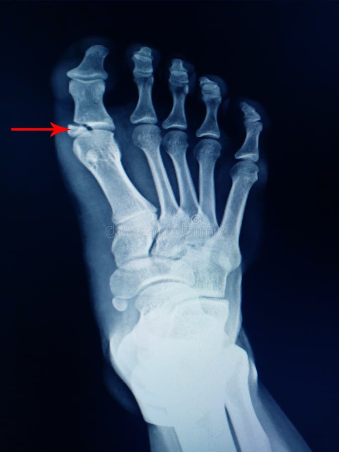

left foot x ray
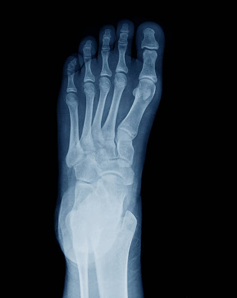
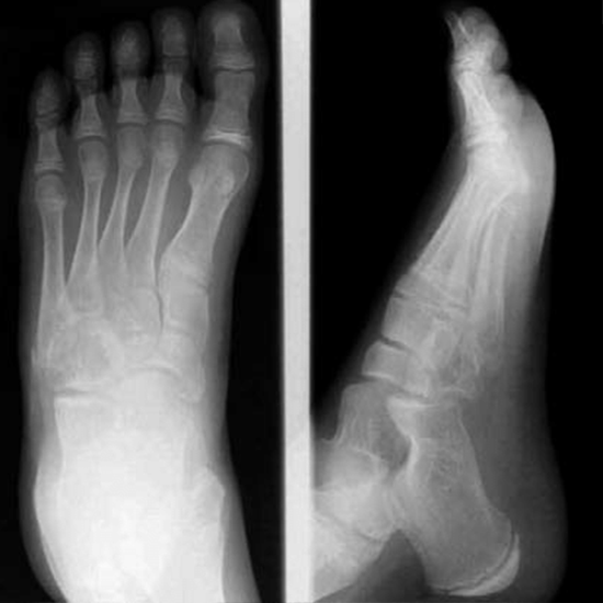
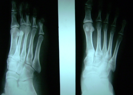
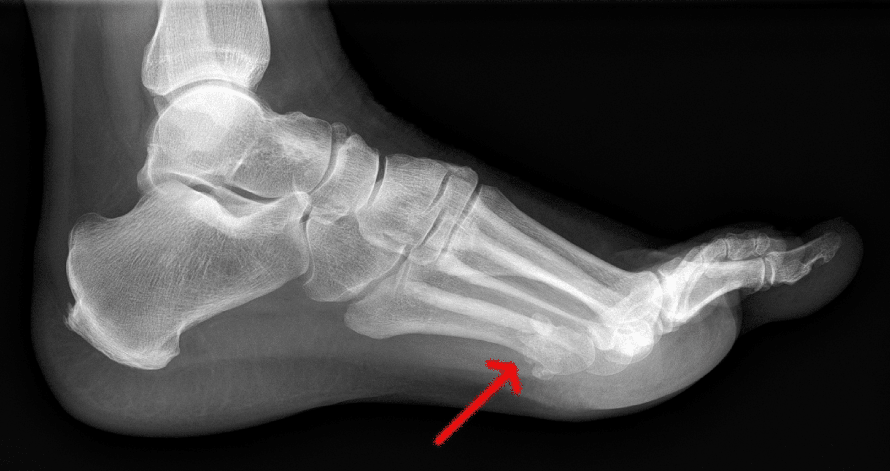
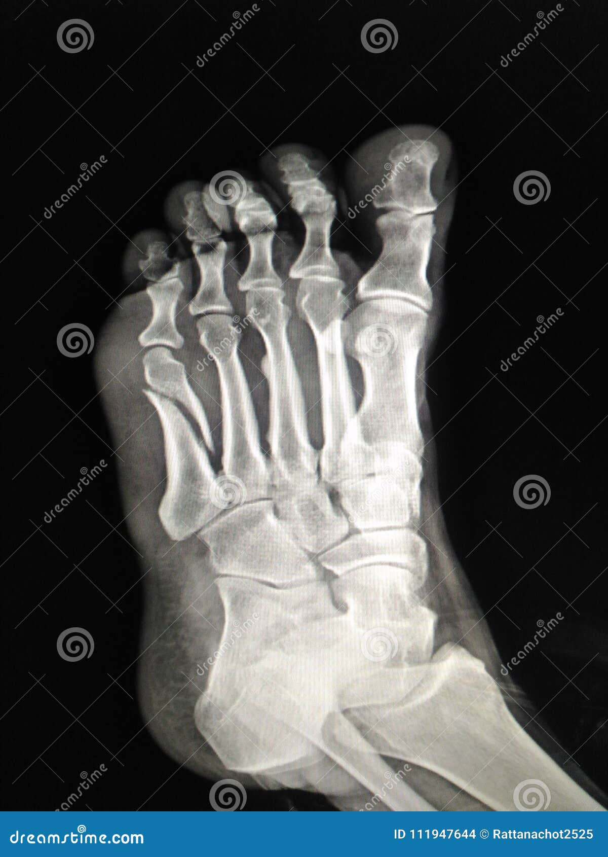
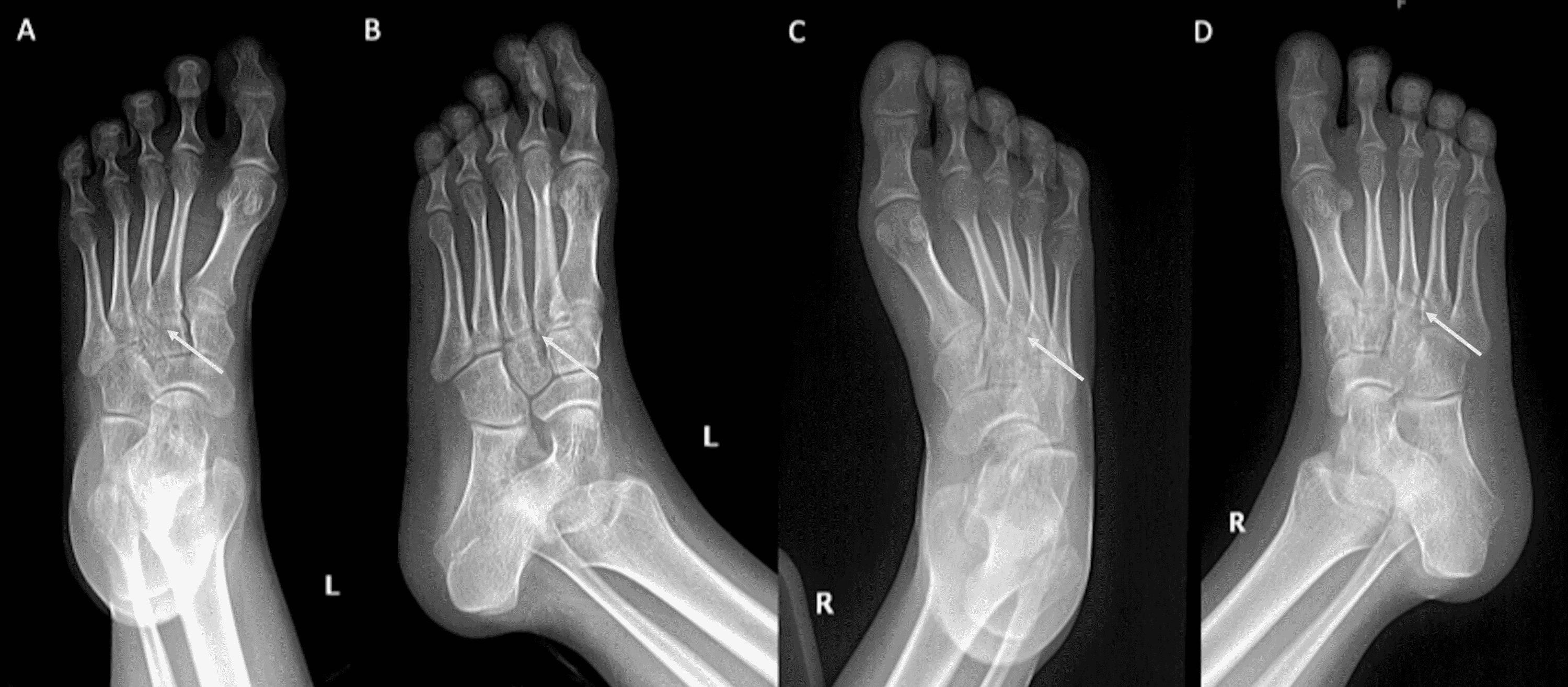
Chụp X-quang chân trái là một trong những phương pháp không xâm lấn phổ biến để chẩn đoán các vấn đề về xương và sụn ở chân. Phương pháp này có thể phân loại thành các loại như: chụp X-quang chân trái bình thường, chụp X-quang chân trái với chấn thương hoặc gãy xương, chụp X-quang chân trái bị viêm, bị phù lên và chụp X-quang chân trái sau khi thực hiện phẫu thuật.
Công dụng của chụp X-quang chân trái
Việc chụp X-quang chân trái được sử dụng để đánh giá sự tổn thương trên chân như gãy, chấn thương hoặc phù lên. Ngoài ra, phương pháp này còn hỗ trợ chẩn đoán các bệnh về xương, nang gan, hoặc các bệnh về hệ thống tự miễn…
Chuẩn bị trước khi chụp X-quang chân trái
Không có yêu cầu đặc biệt nào để chuẩn bị trước khi chụp X-quang chân trái. Tuy nhiên, bạn sẽ cần tháo trang sức hoặc quần áo bị che phủ lên khu vực sẽ được xét nghiệm.
Nếu bạn có thai hoặc cho con bú, bạn nên thông báo cho nhân viên y tế để được tư vấn về những nguy cơ và rủi ro khi thực hiện chụp X-quang chân trái.
Hướng dẫn thực hiện chụp X-quang chân trái
Khi bắt đầu thực hiện chụp X-quang chân trái, bạn sẽ được yêu cầu để đứng hoặc nằm trên giường. Các nhân viên y tế sẽ sắp xếp chân của bạn để thu được hình ảnh chính xác nhất.
Trong khi chụp, bạn sẽ cần giữ chân ở vị trí vô độ trong một khoảng thời gian rất ngắn. Quá trình này không gây đau hoặc khó chịu.
Những rủi ro khi sử dụng chụp X-quang chân trái
Chụp X-quang chân trái được coi là một phương pháp an toàn và không đáng lo ngại. Điều này đặc biệt đúng nếu bạn chỉ thực hiện chụp X-quang định kỳ mà không có các triệu chứng bất thường nào.
Tuy nhiên, nếu bạn cho rằng mình có thai, hoặc đang cho con bú, bạn nên thông báo cho nhân viên y tế để được tư vấn về những nguy cơ và rủi ro khi thực hiện chụp X-quang chân trái.
Các hình ảnh chụp X-quang chân trái bình thường cho nam giới, trẻ em và nữ giới
Hình ảnh chụp X-quang chân trái bình thường cho nam giới:
Hình ảnh chụp X-quang chân trái bình thường cho trẻ em:
Hình ảnh chụp X-quang chân trái bình thường cho nữ giới:
Các hình ảnh chụp X-quang chân trái với gãy xương hoặc chấn thương
Hình ảnh chụp X-quang chân trái với gãy xương hoặc chấn thương:
Các hình ảnh chụp X-quang chân trái bị viêm, bị phù lên và sau khi thực hiện phẫu thuật
Hình ảnh chụp X-quang chân trái bị viêm:
Hình ảnh chụp X-quang chân trái bị phù lên:
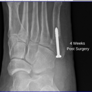
Hình ảnh chụp X-quang chân trái sau khi phẫu thuật:
Thông thường, các chuyên gia sẽ thực hiện chụp X-quang chân trái để chẩn đoán các vấn đề về xương, sụn và các vấn đề khác liên quan đến chân. Phương pháp này là một cách an toàn để giúp bác sĩ chẩn đoán bệnh của bạn và đưa ra liệu pháp điều trị phù hợp. Nếu bạn tin rằng mình đang có vấn đề về chân, bạn nên tham khảo ý kiến của bác sĩ để đưa ra đánh giá chính xác nhất.
Từ khoá người dùng tìm kiếm: left foot x ray normal left foot x ray male, normal left foot x ray child, normal left foot x ray female, normal foot x ray vs fracture, foot x-ray images normal, right leg foot x ray, normal left foot oblique x ray, abnormal foot x ray
Tag: Update 66 – left foot x ray
Anatomy of Foot X-rays
Xem thêm tại đây: huanluyenchosaigon125.com
Link bài viết: left foot x ray.
Xem thêm thông tin về chủ đề left foot x ray.
- 593 Left Foot Xray Images, Stock Photos & Vectors | Shutterstock
- Left foot X-ray: (a) Anteroposterior view; (b) lateral view; (c)…
- Normal foot x-ray: MedlinePlus Medical Encyclopedia Image
- Normal foot x-rays | Radiology Case | Radiopaedia.org
- X-ray Left Foot AP/LAT/Oblique | Test Price in Delhi
- Foot xray hi-res stock photography and images – Alamy
Categories: blog https://huanluyenchosaigon125.com/img
